My Role
Responsible for research, planning, wireframing, high fidelity design, prototyping, user feedback, grooming
THE TEAM
1 Design consultant, 2 Designers, 2 Product managers
PRODUCT OVERVIEW
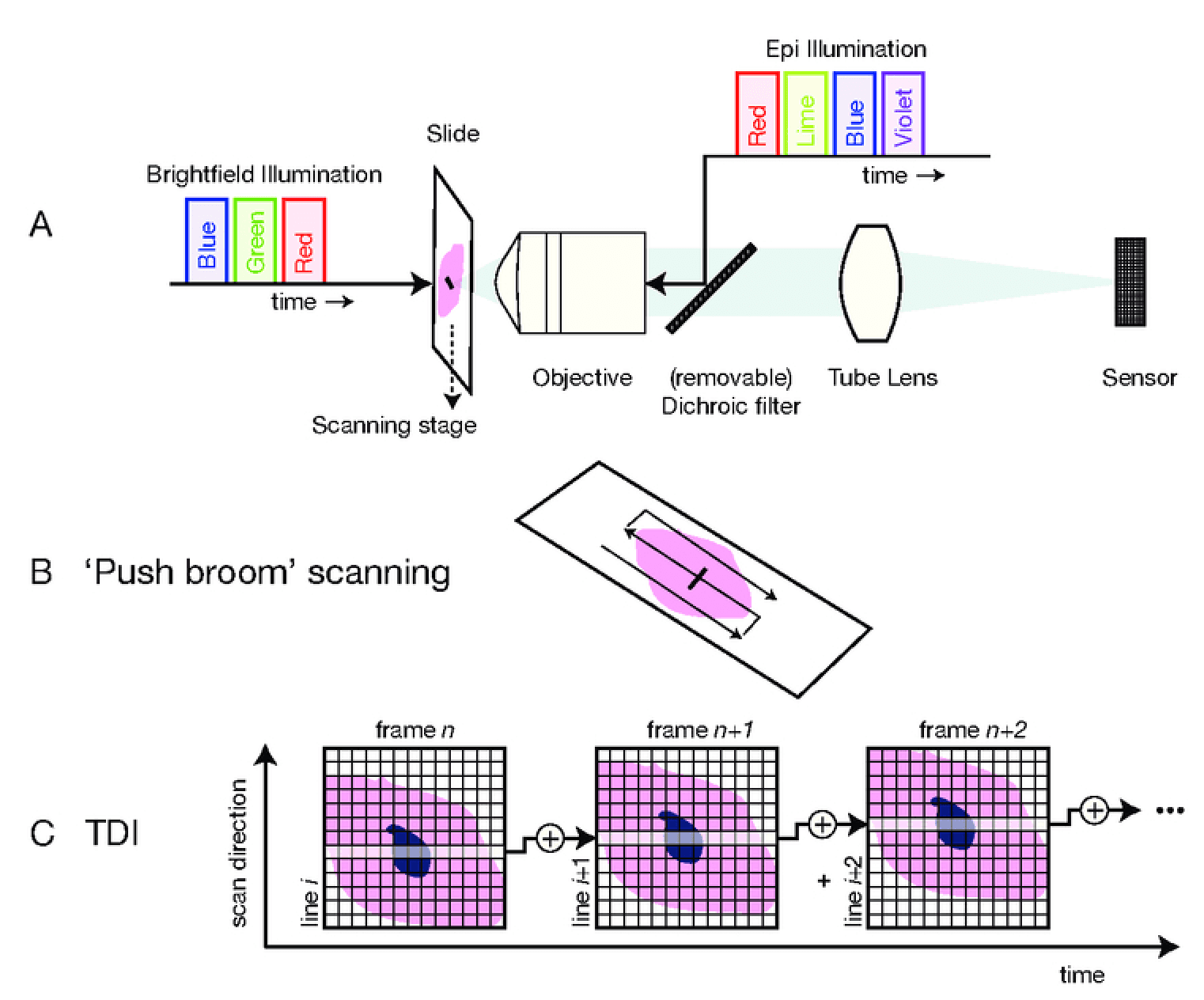
Pathology is a branch of medical science that involves the examination of tissues, organs, bodily fluids, and autopsies in order to study and diagnose a disease. Digital pathology refers to the digitization of conventional pathology lab and tests in order to increase workflow efficiency via faster and accurate analyses. Although diagnosis using manual microscopic review is still a gold standard in many areas, there is also a huge demand for automation of pathology systems. Automated digital microscopy systems, also known as whole slide imaging (WSI) systems, partially automate the process of review. They capture digital images of the physical slide and create a “virtual slide”. This virtual slide can then be reviewed by multiple experts, enabling both remote review and collaborative review, and also opens up the possibility of automated analysis by artificial intelligence (AI) systems. It has many advantages such as scanning of slides with ideal focus, multiple magnifications and developing enhanced resolution digital images in a comparatively shorter time span. This has led to a number of new opportunities not possible using conventional microscopes, including digital collaboration/telepathology, integration with electronic workflows and health records, and diagnostic support based on computational tools like artificial intelligence.

Target audience
The biggest need for a WSI system is in primary care, especially in non urban areas of developing countries, where there is a lack of trained clinical professionals.
Pain points
Issues in manual microscopy/current workflow from product perspective:
Difficulty in observing low contrast samples: Sample details cannot be clearly observed with low contrast and low reflective difference even after magnification.
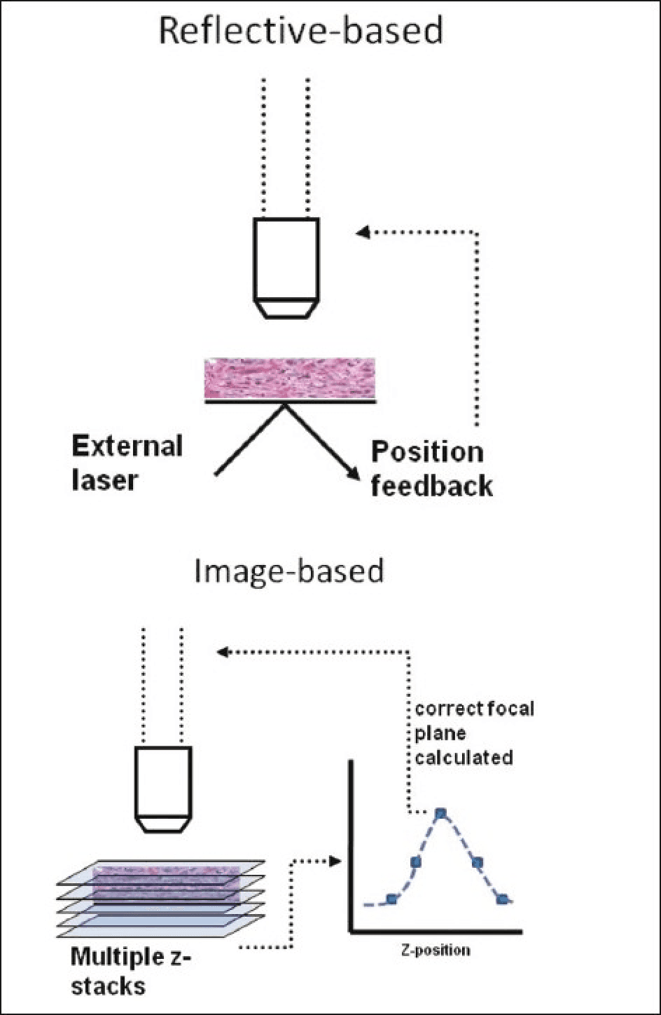
Focusing issues : Areas of an uneven sample surface are hard to view in clear focus at the same time and manual focusing can be erroneous.
High TAT - Longer turnaround time because of manual workflow.
Requires pathologists to be co-located : Increases TAT for tests as Pathologists need to be present at the lab to analyze the sample.
Financial constraints : The financial constraints that come with lab testing hampers the quality of pathology.
Difficulty in storing/transporting samples : The integrity of samples could be compromised if they are not kept in consistent conditions until they are reviewed.
Issues in maintaining patient records : All the samples/reports of a particular case need to be physically stored if they need to be referred later.
User Research Perspective:
1. Pathologists:
- Inadequate image resolution and quality: Pathologists require high-resolution and high-quality images to accurately analyze and diagnose slides. The design should prioritize advanced imaging hardware and algorithms to ensure clear and detailed representations of histopathological slides.
- Limited annotation capabilities: Pathologists need comprehensive annotation tools to mark, highlight, and annotate regions of interest. The design should incorporate intuitive and versatile annotation features, including shapes, colors, transparency options, and the ability to add textual annotations.
- Cumbersome navigation and image exploration: Pathologists face challenges with complex user interfaces and inefficient navigation features. The design should prioritize an intuitive user interface with smooth navigation, zooming, and panning functionalities to facilitate easy exploration of digital slides.
2. Researchers:
- Limited analysis tools and algorithms: Researchers require advanced analysis capabilities, including quantitative assessment, pattern recognition, and image processing. The design should incorporate powerful analysis tools and algorithms to enable in-depth research and meaningful insights.
- Integration with research software: Researchers often work with specific research software and tools. The design should ensure compatibility and seamless integration with existing research software, allowing for efficient data exchange and collaboration.
- Data management capabilities: Researchers handle large volumes of digital slide data, necessitating efficient data management features. The design should provide robust data organization, search, and retrieval functionalities to streamline research workflows.
3. Clinicians:
- Integration with electronic health record (EHR) systems: Clinicians require seamless integration between WSI tools and EHR systems to access patient data and correlate digital slide findings with clinical information. The design should prioritize interoperability with EHR systems to enable efficient clinical decision-making and patient management.
- Quick image loading and processing: Clinicians often need rapid access to digital slides for immediate analysis. The design should optimize image loading and processing speed to support fast and responsive workflows.
- Collaboration and consultation capabilities: Clinicians rely on effective collaboration and consultation for patient care. The design should facilitate easy sharing of digital slides with colleagues or specialists, enabling remote collaboration, second opinions, and consultations.
Issues in existing WSI systems
High cost - Considering the advancements in performance and reduction in cost of both compute systems and camera systems, the cost of state-of-the-art WSI systems still remains high.
Inaccurate & Inefficient - Current automated focusing algorithms, especially those deployed in cost effective microscopy systems, often cannot match the efficiency of a skilled human operator in keeping a sample in focus
Rarely used - WSI systems are typically used in large tertiary care clinical laboratories or research institutions only.
Problem statement
The problem lies in the inadequate accuracy and advanced features of existing whole slide imaging platforms, hindering efficient annotation and analysis of digital slides, and impeding collaboration in image analysis workflows. The lack of optimized workflows further exacerbates these challenges and limits integration with digital pathology systems. To address these issues, the design of a high-accuracy platform must prioritize the integration of robust annotation tools and workflow optimization. This includes comprehensive annotation features and smart capabilities to enhance efficiency in collaborative environment.
Our Vision
WSI system is intended to be the base product which can be used by customer to scan different kinds of specimen enabling the customer to use benefits of Digital Pathology like remote review, reduced TAT and ease of maintaining samples. Our goal is to make WSI systems affodable and accessible to even remotes areas where there is lack of skilled pathologists. We are targeting the tests with critical TAT, microscopic based tests requiring longer sample storage and tests requiring second opinion.
Our Competitors
WSI system is intended to be the base product which can be used by customer to scan different kinds of specimen enabling the customer to use benefits of Digital Pathology like remote review, reduced TAT and ease of maintaining samples. We are targeting the tests with critical TAT, microscopic based tests requiring longer sample storage and tests requiring second opinion.
Features
Morphle Labs
CA Microsope
Easy Zoom
Scale
Freehand Drawing to mark a section and to automatically calculate areas
Define a bounding area and auto-compute area
Measure Length
Rotate Left
Rotate Right
Snapshot of Screen
Map of section under view
Create Annotations
View Annotations List
Pin Annotations
Annotation Tags
Layer Management
Full Screen
Side-By-Side Viewer
Share Images as links or embeddable iframes
Segment - This tool allows you to display, count, and export nuclear segmentations
on the image. Clicking this tool opens the following custom toolbar.
Add Custom Models (Tensor-Flow Compatible)
Proposed solution
The proposed solution is to design a Whole Slide Imaging (WSI) platform that addresses the identified user challenges through the following key features:
High-resolution imaging
Comprehensive annotation tools
User-friendly interface and navigation
Advanced analysis capabilities
Seamless integration with research software
Efficient data management
Collaboration and consultation features
Flows and wireframing
We started with low-fidelity design with the flows provided by product team, and came up with rough concepts to share with larger team and discuss about the approach. After a lot of back and forth we were able to decide on the approach we thought was correct. We decided to verify it by talking to potential users.
user feedback
We have received a lot of feedback about how people perceive and use the computer application versus a regular manual microscope. Since they will be performing the same tasks on the digital platform, it was really important for us to make sure it works as closely as possible to the manual microscope in terms of how it functions and how accurate it is.
Whole Slide Imaging (WSI)
Research | Planning | Designing